The inverted microscope is equipped with a Zeiss Airyscan 2 detector, which enables high-resolution imaging with any fluorescently labeled sample. The multiplex mode offers up to 8 times faster and more gentle imaging compared to conventional confocal microscopy, which is ideal for long-term imaging of living cells. Focus stabilization is achieved by the Definite Focus system.
The microscope is fully enclosed with heating and CO2 environmental control and is equipped with a motorized stage.
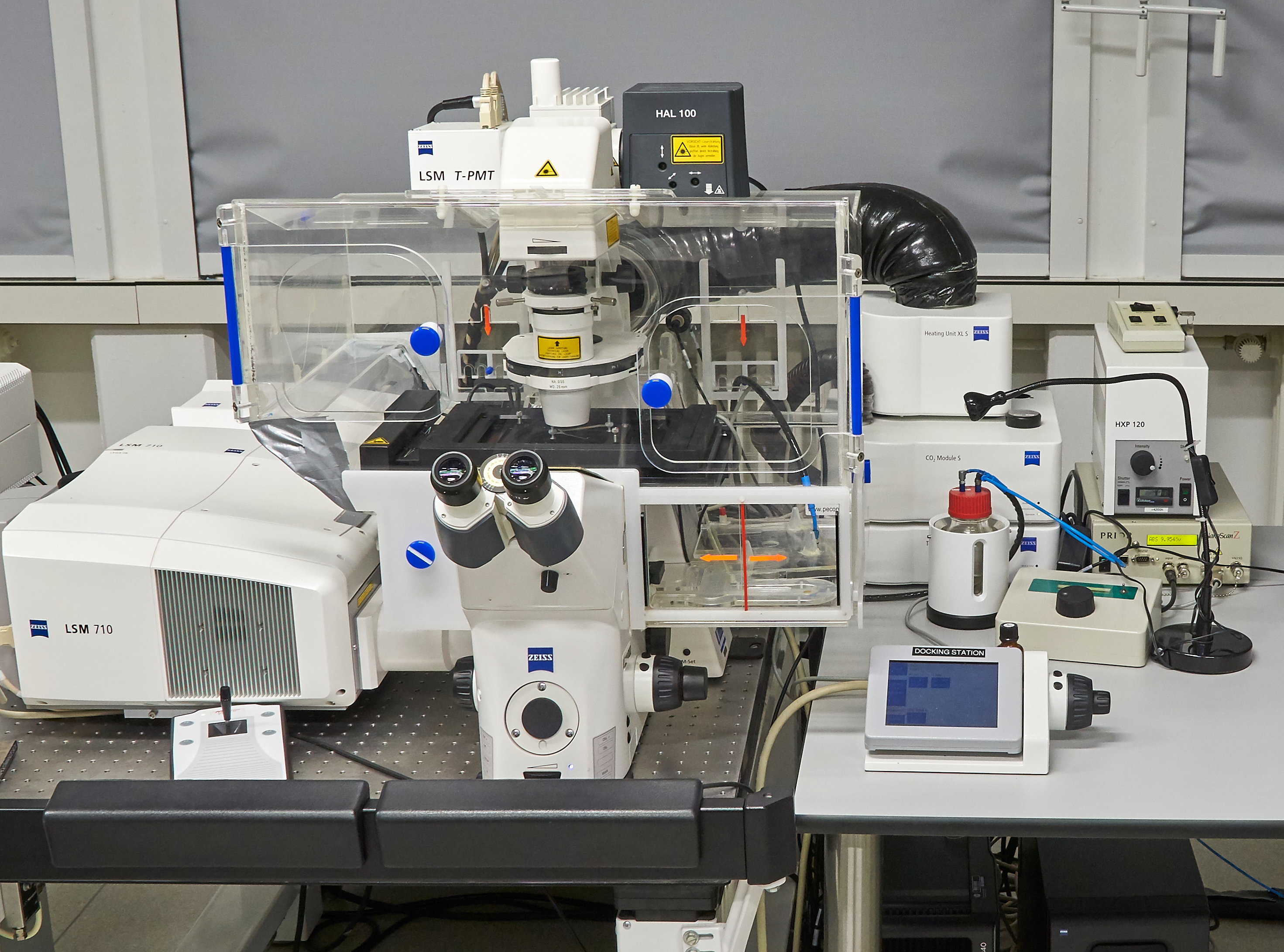
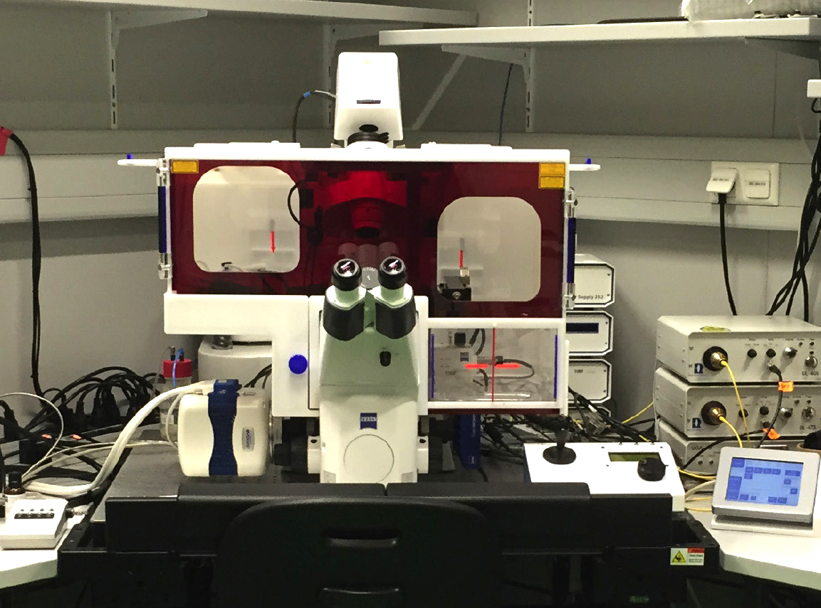
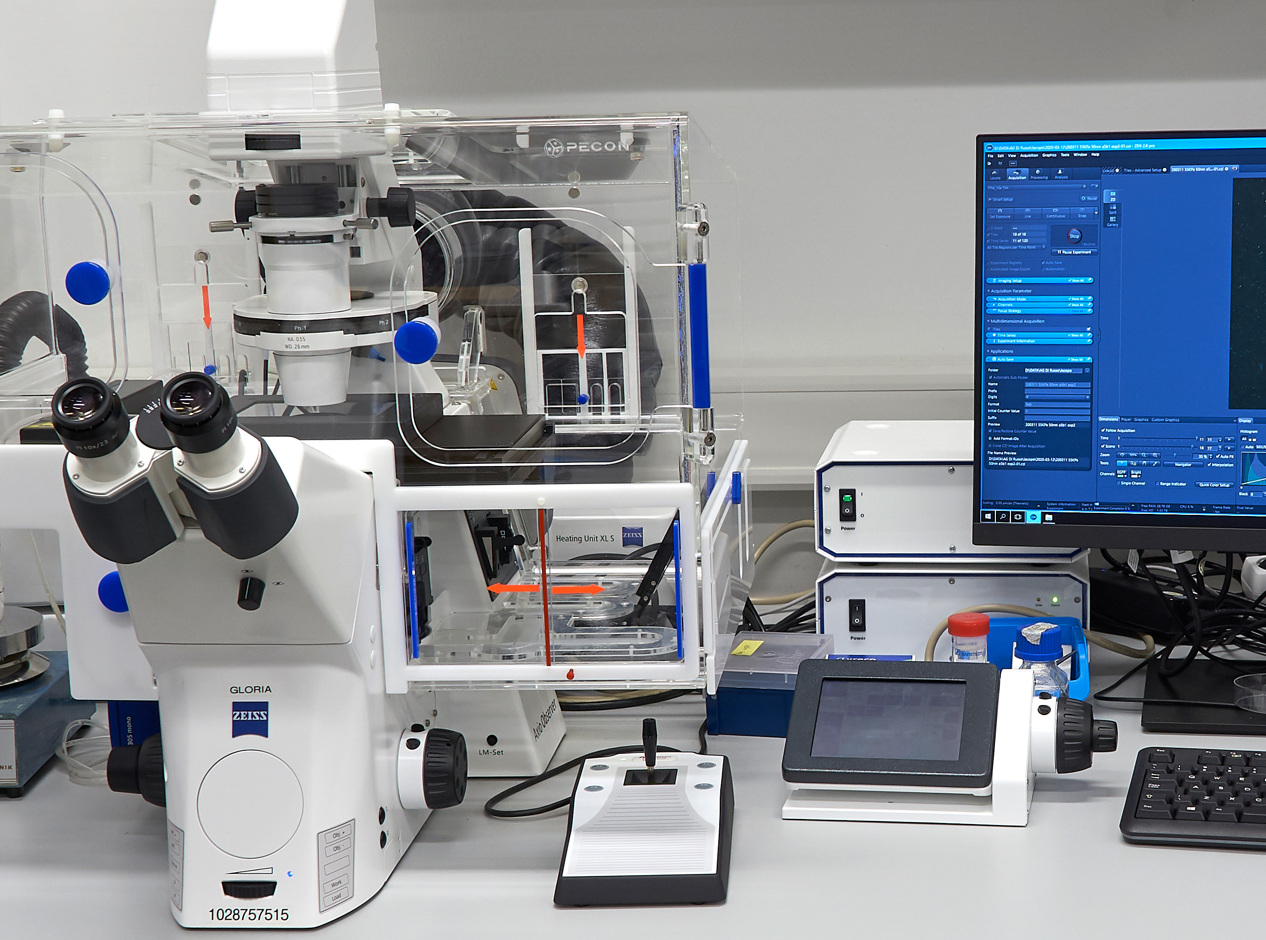
This Zeiss LSM 710 confocal system uses a Zeiss AXIO Observer Z1 inverted microscope stand with transmitted light illumination a light source for fluorescence excitation HXP 120 and laser illumination sources. It can collect transmitted light images (bright field and DIC) as well as conventional and confocal fluorescence images and has superresolution capabilities (AIRYSCAN). The microscope is fully enclosed for heating and CO2 environmental control and is equipped with a motorized stage. Wide array of imaging applications including protein localization analysis, live cell imaging of cells and animals.
The system comes with a Zeiss AXIO Observer Z1 inverted microscope stand with transmitted light illumination a light source for fluorescence excitation HXP 120 and laser illumination sources. It can collect transmitted light images (bright field and DIC) as well as conventional and confocal fluorescence images. The microscope is fully enclosed for heating and CO2 environmental control and is equipped with a motorized stage.
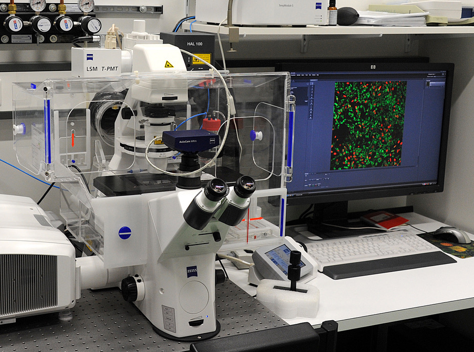
This Zeiss LSM 710 can be used for the generation of confocal images of fixed samples and the investigation of living cells.
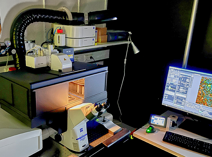
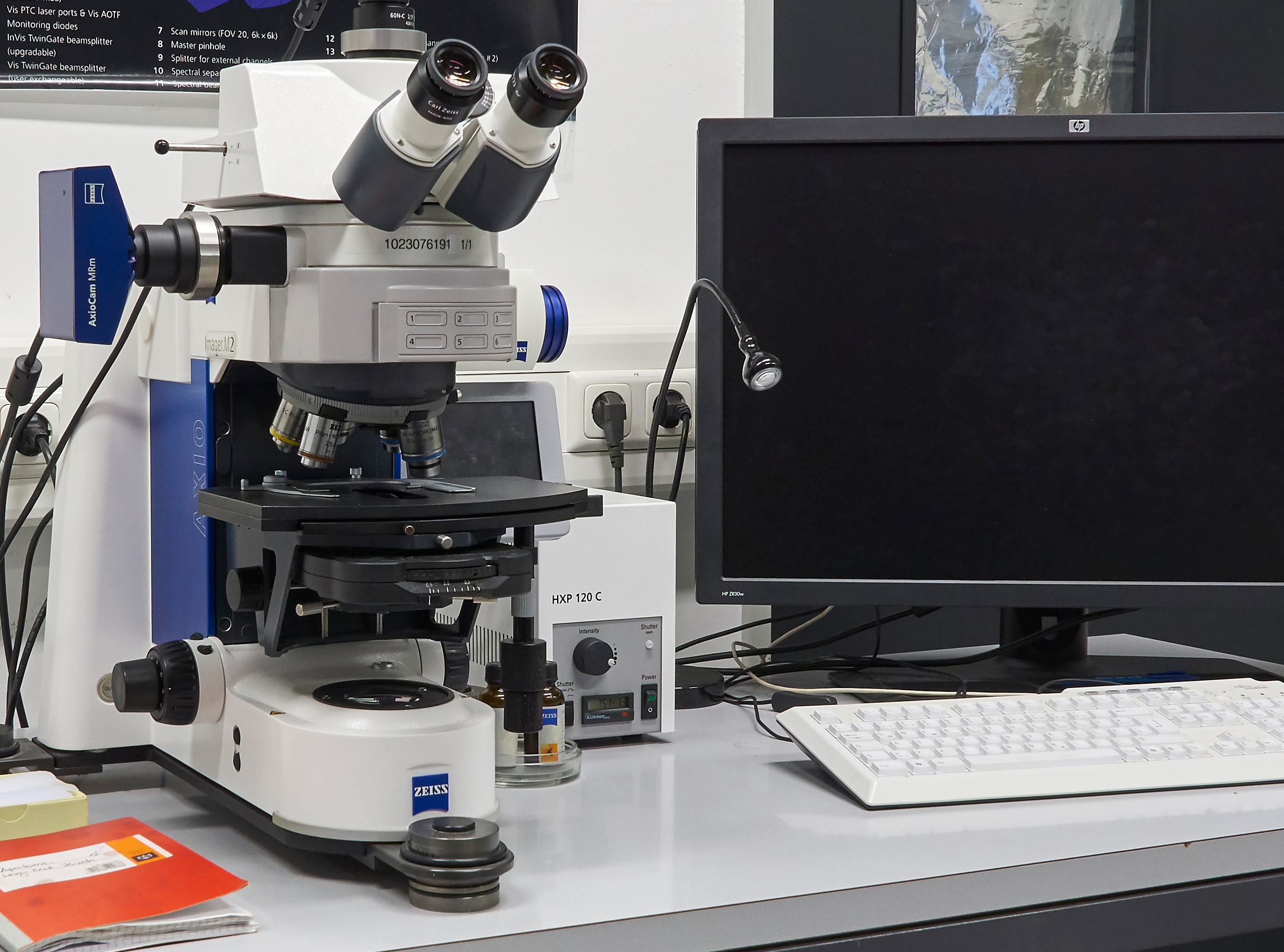
This Zeiss LSM 700 confocal system uses a Zeiss AXIO Observer inverted microscope stand with transmitted light illumination, a light source for fluorescence excitation HXP 120 and laser illumination sources. It can collect transmitted light images and confocal fluorescence images. It is a basic confocal microscope, useful for standard imaging techniques.
more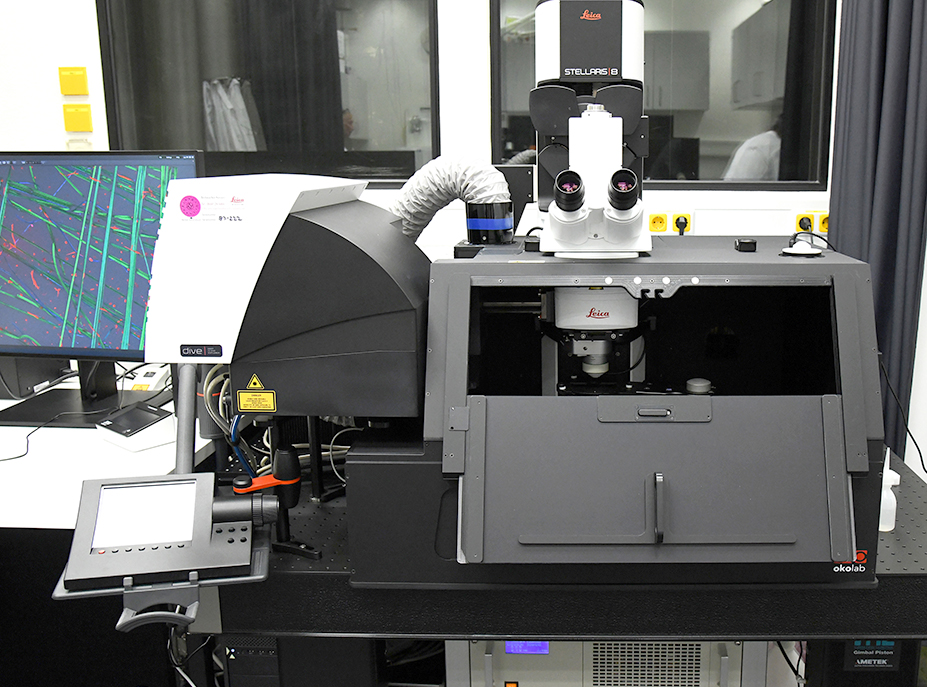
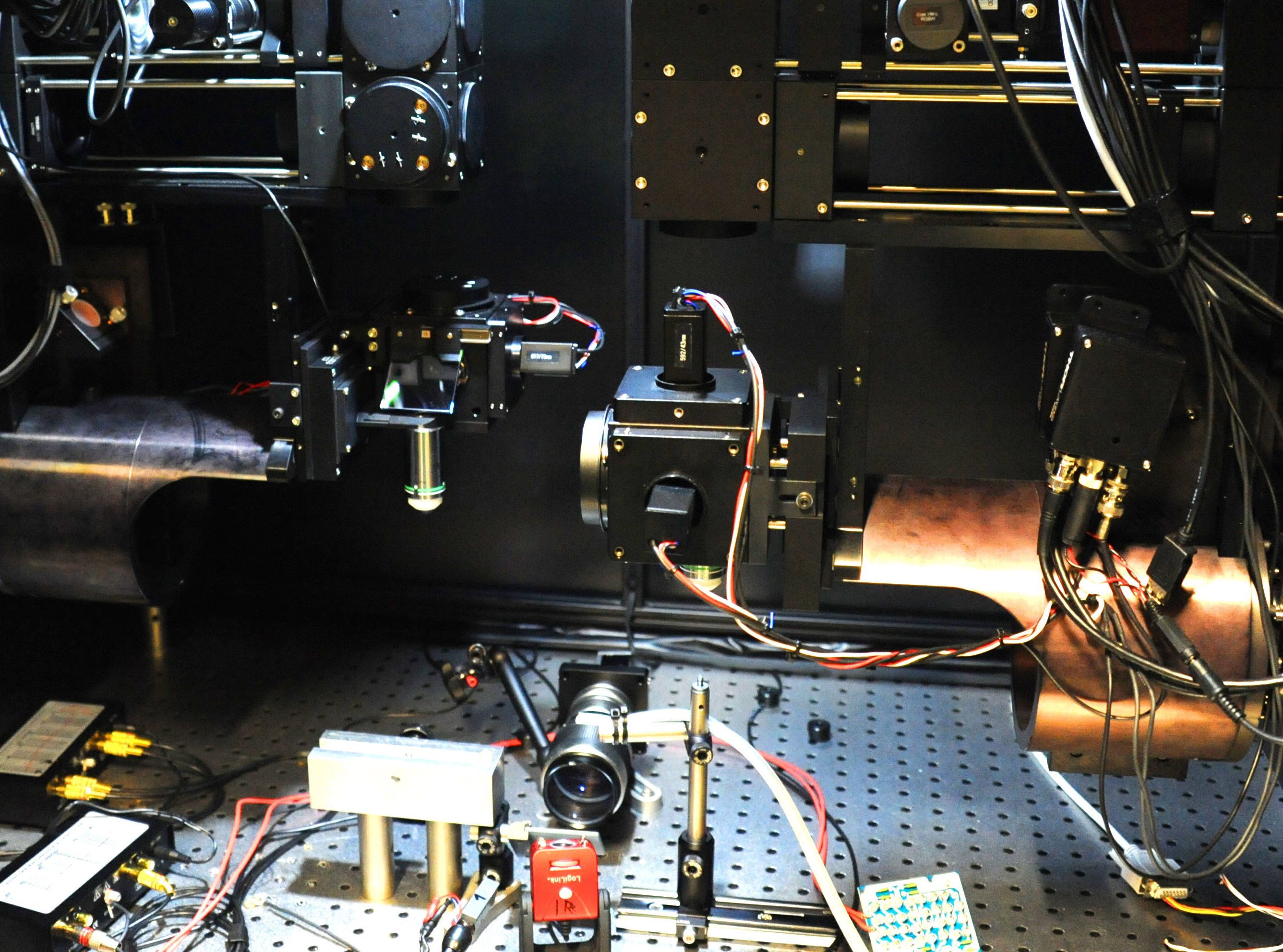
The system enables fluorescence excitation with a dual line laser in the infrared range, multiplexing using spectral information and video-rate FLIM. Optional beam routing to achieve a balance between resolution and penetration depth. The InSight X3 Laser covers a broad excitation range from 680 nm to 1300 nm for optimal penetration, imaging of far red probes, three-photon excitation, and third harmonic generation imaging. The single beam can be combined with the fixed 1045 nm dual beam option. This supports the diverse needs of multimodal imaging.
more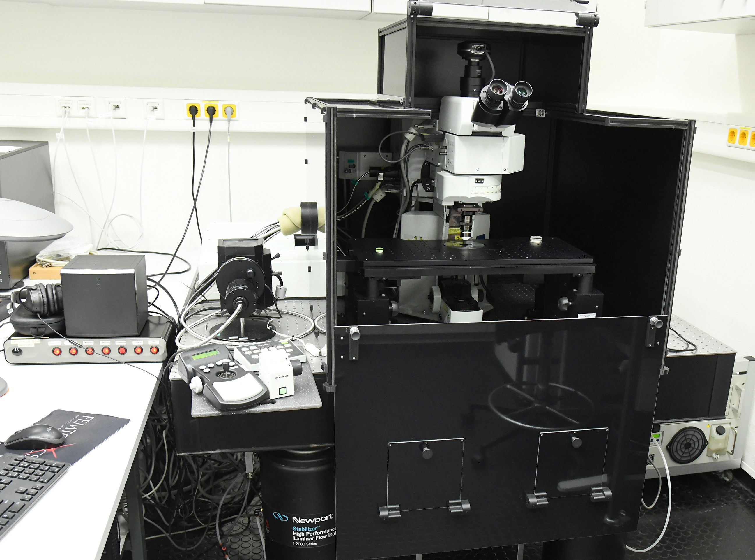
Supports multicolor fluorescent studies for imaging of living, whole mount or thickly sliced specimens. Dynamic biological processes can be imaged hundreds of micrometers within living cells and tissues. Provides support for applications where phototoxicity/photobleaching are a concern such as time course studies of living cells and tissues. Low magnification lens and long working distance stage allow imaging of large samples, embryos, and animals. A range of microprobe objectives are available for minimally invasive in vivo imaging of deep tissues. This microscope also offers conventional confocal laser scanning of samples on slide.
The custom design ensures fast and reproducible access to the area of interest, including rotation of imaging plane, and maximizes space for auxiliary experimental equipment in the vicinity of the animal. Mechanical flexibility is achieved with large motorized linear stages that move the objective in the X, Y, and Z directions up to 130 mm. +- 45° rotation of the frontend (rotational freedom for one axis) is achieved with the combination of a motorized high precision bearing and gearing. The high-speed two-photon microscope is able to scan large volumes at high speeds. Resonant scanning technology is used to produce two-dimensional (x,y) images with high frame rates (512x512 pixels at 30 Hz). A tuneable lens system is used to quickly shift the focus in z dimension. The combination of these two systems is able to scan 3D volumes in-vivo (1 x 1 x 0.5mm at 8 Hz).
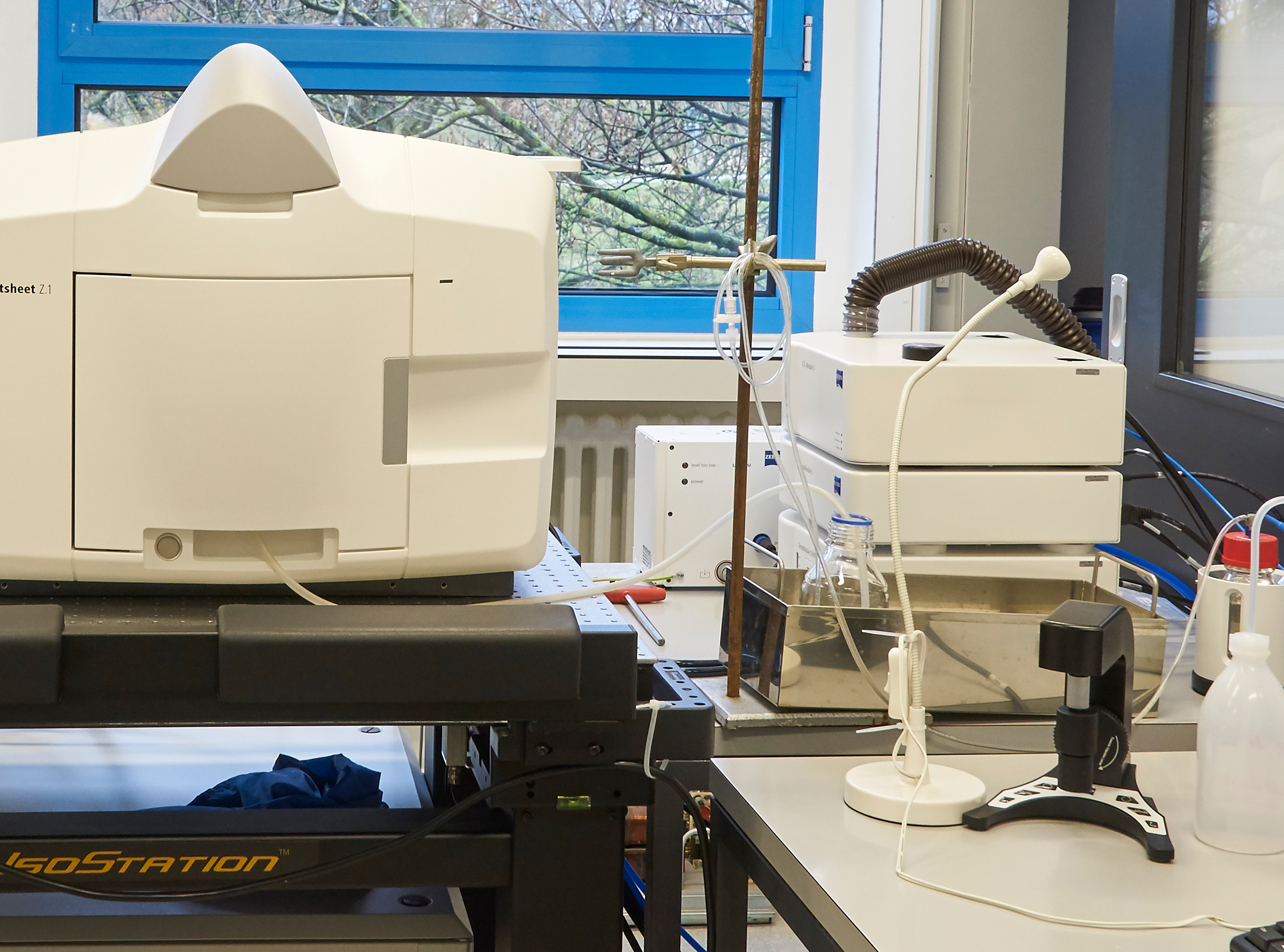
The Multiview Light Sheet Fluorescence Microscope performs fluorescence imaging on large, living samples, with low phototoxicity or bleaching and with high temporal resolution. Lightsheet Z.1 allows researchers to record the development of large, living samples. It acquires images of the whole sample volume at sub-cellular resolution - in a fraction of the time it takes using other techniques.
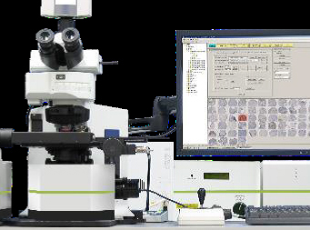
The Vectra® 3 is a powerful system merging automated slide-handling, multispectral imaging technology, and unique pattern-recognition-based image analysis in one automated workflow. This system accurately measures protein expressions and morphometric characteristics in distinct tissue regions of interest or on whole slides. Tissue sections or TMAs can be labeled with immunofluorescent (IF) or immunohistochemical (IHC) stains, or with conventional stains (e.g., H&E). When using IF or IHC stains, multiple proteins can be measured on a per tissue, per cell, or per cell compartment (e.g. nuclear, cytoplasmic) basis - even when signals are spectrally similar, are located in the same cellular compartment or are obscured by autofluorescence.
The TIRF 3 system is based on the Axio Observer Z1 inverted microscope. It is equipped with HXP-120 and lasers for fluorescence microscopy. Several filter sets allows multi-channel fluorescence microscopy for imaging of either fixed or living cells. Environmental control is achieved by regulation of temperature and carbon dioxide. The UGA-40 FRAP unit is driven via TTL with Zen 2 (blue) and allows single or multiple FRAP experiments.
The upright Zeiss Imager ApoTome 2 optical sectioning microscope is a state of the art fluorescence microscope that additionally can be operated using structured illumination. In this mode it creates optical sections of fluorescent samples free of scattered light. The system calculates your optical section from three images with different grid positions to prevent scattered out-of-focus light.
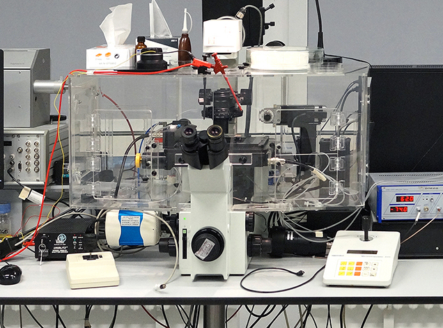
The Olympus IX70 life cell microscope is inverted fluorescence microscope that is aimed for the application of various life cell imaging setups. It is specially equipped with a monochromator, a motorized stage, an incubation chamber and two micromanipulators.

The MULTIZOOM AZ100M offers the combined advantages of a stereoscopic microscope with a wide field of view and long working distance, and epifluorescence illumination. It offers a magnification range from 5x to 400x.
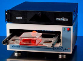
Gain insights into biological processes of cells in real time via non-perturbing quantitative analysis IncuCyte® ZOOM System enables observation and quantification of cell behavior over time by automatically gathering and analyzing images around the clock within a standard incubator.
Each IncuCyte® ZOOM System houses multiple T-flasks or microtiter plates (up to 6) and can acquire >2000 images per hour.

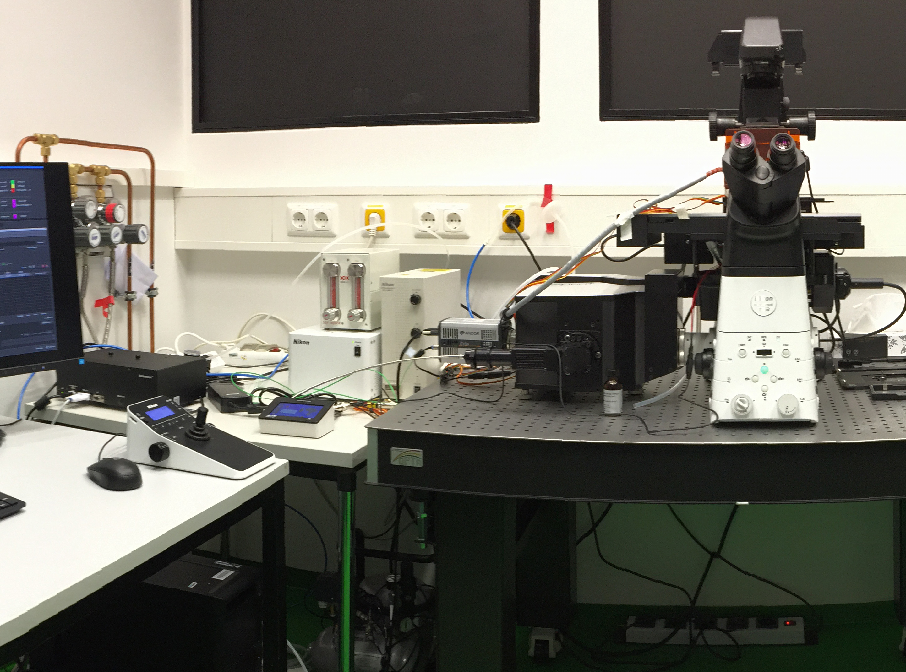
The microscope Nikon Ti2-E with Crest X-Light V2 Spinning Disk is very flexible inverted fluorescence platform suited to live cell image time-lapse microscopy either in confocal or fluorescence widefield mode. Allowing high resolution TLM or medium/high-throughput phenotypic screenings. The environmental control is achieved by an IBIDI incubation system with CO2 mixer compatible with one to four 8well u-Slices and 35mm u-Dishes.
The microscope Nikon Ti2-E with Crest X-Light V2 Spinning Disk is very flexible inverted fluorescence platform suited to live cell image time-lapse microscopy either in confocal or fluorescence widefield mode. Allowing high resolution TLM or medium/high-throughput phenotypic screenings. The environmental control is achieved by an IBIDI incubation system with CO2 mixer compatible with one to four 8well u-Slices and 35mm u-Dishes.
Axio Observer is an inverted fluorescence microscope suited for demanding multimodal imaging of living and fixed specimens. It is equipped with motorized stage and automated reflectors and objective turrets. Light illumination is provided by LED.