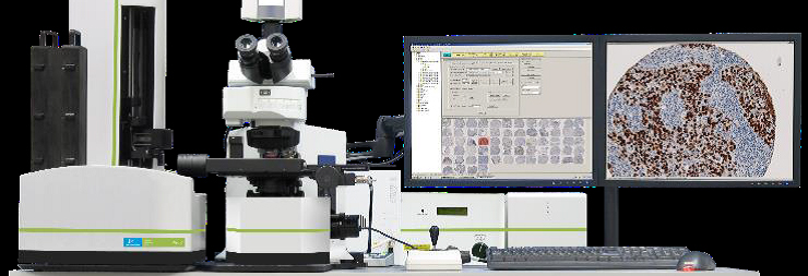
PerkinElmer Vectra 3.0 Automated Quantitative Pathology Imaging System
The Vectra® 3 is a powerful system merging automated slide-handling, multispectral imaging technology, and unique pattern-recognition-based image analysis in one automated workflow. This system accurately measures protein expressions and morphometric characteristics in distinct tissue regions of interest or on whole slides. Tissue sections or TMAs can be labeled with immunofluorescent (IF) or immunohistochemical (IHC) stains, or with conventional stains (e.g., H&E). When using IF or IHC stains, multiple proteins can be measured on a per tissue, per cell, or per cell compartment (e.g. nuclear, cytoplasmic) basis - even when signals are spectrally similar, are located in the same cellular compartment or are obscured by autofluorescence.
Applications:
Objectives |
|||
magnif. |
NA |
WD |
type |
4 x |
0.16 |
13 mm |
Universal Plan Super Apochromat |
20 x |
20 x |
0.6 mm |
Universal Plan Super Apochromat |
40 x |
0.95 |
0.18 mm |
Universal Plan Super Apochromat |
|
|||
|
|||
|
Microscopy: |
|
|
Computer: |
|
|
Software: |
Vectra® software controls the system. Inform® software analyzes PerkinElmer Vectra multispectral images and uses a combination of machine-learning, pattern-recognition-based tissue segmentation and multispectral imaging for automated image analysis and extraction of signals from tissue markers. Allows for quantitative analysis of biomarker expression in tissue sections and TMAs, separation of weakly expressing and overlapping markers, cellular analysis of H&E, IHC and immunofluorescence in fixed tissues, and automatic identification of specific tissue types using trainable feature recognition algorithms. The study workflow interface supports batch processing, rapid segmentation review, and data merging. Capable of exporting spectrally unmixed signals on a per-cell basis from within selected tissue-segmentation areas. Phenochart® software enables viewing and annotation of Brightfield and Fluorescent slides scanned by PerkinElmer’s Vectra 3 Automated Quantitative Pathology Imaging System. |