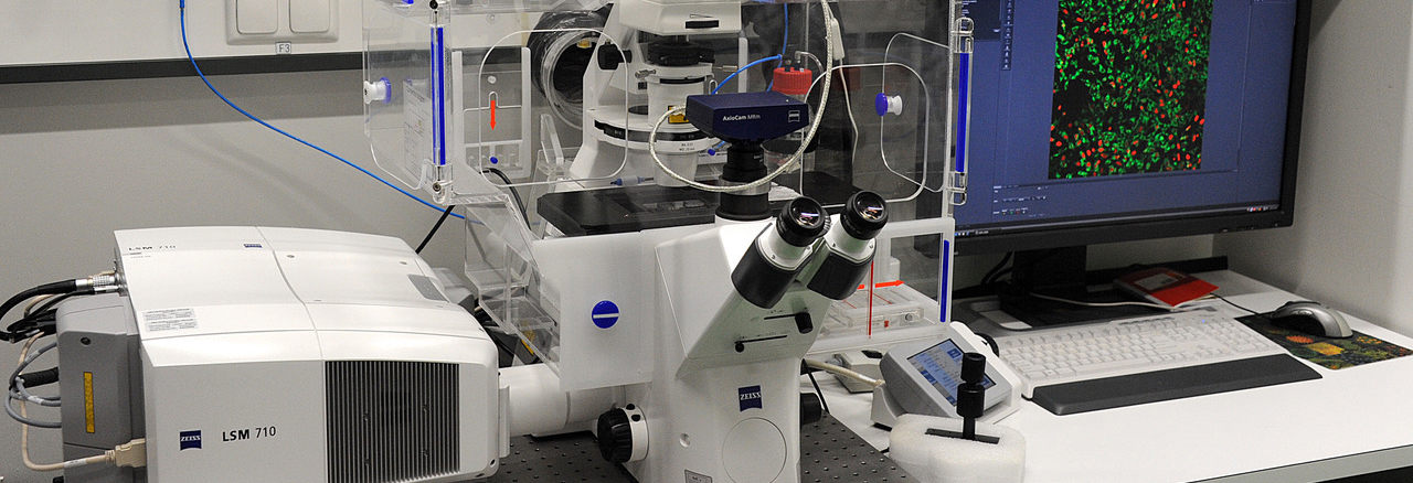
Inverted Zeiss LSM 710 Laser Scanning Confocal Microscope with 34-Channel Quasar Detector
The system comes with a Zeiss AXIO Observer Z1 inverted microscope stand with transmitted light illumination, light source for fluorescence excitationing HXP 120 and laser illumination sources. It can collect transmitted light images (bright field and DIC) as well as conventional and confocal fluorescence images. The microscope is fully enclosed for heating and CO2 environmental control and is equipped with a motorized stage.
This Zeiss LSM 710 can be used for the generation of confocal images of fixed samples and the investigation of living cells.
The Zeiss LSM710 belongs to the Confocal Microscopy Facility of the IZKF and is located in the University Hospital. It can be booked by all researchers of RWTH Aachen University.
For more information, visit our website at
or contact
Prof. Dr. Gerhard Müller-Newen
gmueller-newen@ukaachen.de
Tel.: +49 241 80 88860
Dr. Sabrina Ernst
sabernst@ukaachen.de Tel.:
+49 241 80 88838
Applications:
Confocal images:
Advanced microscopy:
Objectives |
|||
magnif. |
NA |
type |
specific |
10 x |
0.30 |
Plan-Neofluar |
|
20 x |
0.80 |
Plan-Apochromat |
|
40 x |
1.10 |
C-Apochromat |
Water immersion |
63 x |
1.20 |
C-Apochromat |
Water immersion |
63 x |
1.40 |
Plan-Apochromat |
DIC/Oil immersion |
|
|
Lasers |
|
wavelenght |
type |
405 |
Diode |
458 |
Argon |
488 |
Argon |
514 |
Argon |
561 |
Diode pumped solide state (DPPS) |
633 |
HeNe |
|
|
detectors |
filter or beamsplitter |
Quasar 32-channel array Two PMT detectors |
MBS 405 MBS 445 MBS 458 MBS 458/514 MBS 458/561 MBS 458/514/561/633 MBS 488 MBS 488/561 MBS 488/561/633 T80/R20 |
|
|
Transmitted light detector (TPMT) |
zeiss filter set 38, GFP zeiss filter set 43, Cy3 zeiss filter set 46, YFP zeiss filter set 47, CFP zeiss filter set 49, DAPI |
|
|
Ocular |
zeiss filter set 38, GFP zeiss filter set 43, DsRed zeiss filter set 46, EGFP zeiss filter set 47, CFP zeiss filter set 49, DAPI zeiss filter set 50, Cy5 |