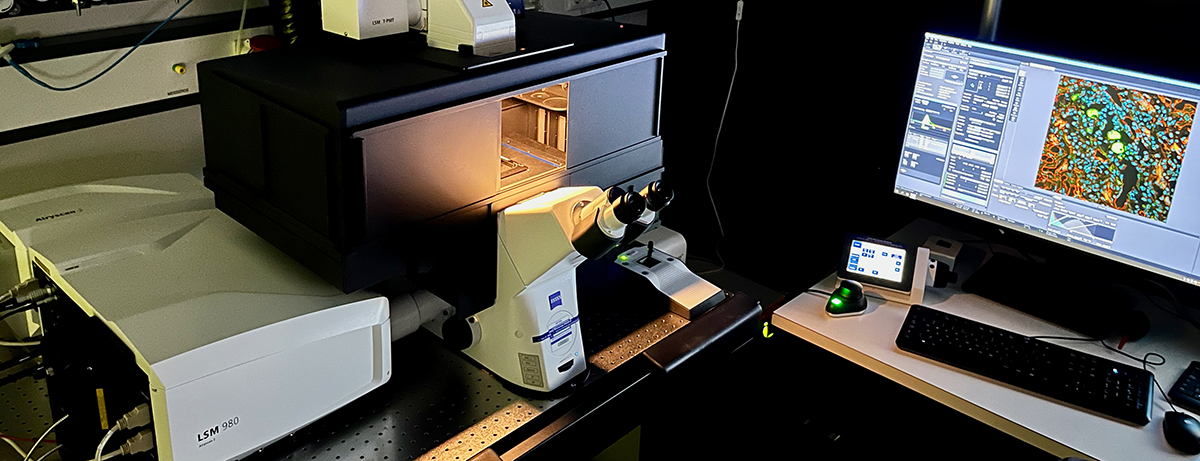
Zeiss LSM 980 with Airyscan 2 confocal laser scanning microscope
The inverted microscope is equipped with a Zeiss Airyscan 2 detector, which enables high-resolution imaging with any fluorescently labeled sample. The multiplex mode offers up to 8 times faster and more gentle imaging compared to conventional confocal microscopy, which is ideal for long-term imaging of living cells. Focus stabilization is achieved by the Definite Focus system.
The microscope is fully enclosed with heating and CO2 environmental control and is equipped with a motorized stage.
The Zeiss LSM 980 with Airyscan 2 belongs to the Confocal Microscopy Facility of the IZKF and is located in the University Hospital. It can be booked by all researchers of RWTH Aachen University.
For more information, visit our website at
or contact
Prof. Dr. Gerhard Müller-Newen
gmueller-newen@ukaachen.de
Tel.: +49 241 80 88860
Dr. Sabrina Ernst
sabernst@ukaachen.de Tel.:
+49 241 80 88838
Applications:
Confocal Microscopy can be used to generate optical slices of fluorescently labeled samples and to examine living cells with advanced fluorescence techniques
Confocal images:
Advanced fluorescence techniques:
Objectives |
|||
magnif. |
NA |
type |
specific |
2.5 x |
0.085 |
EC Plan-Neofluar |
|
10 x |
0.45 |
Plan-Apochromat M27 |
|
25 x |
0.80 |
LD LCI Plan-Apochromat |
Multi immersion |
40 x |
1.20 |
LD LCI Plan-Apochromat |
Multi immersion |
40 x |
1.30 |
Plan-Apochromat |
Oil immersion |
|
|
Lasers |
|
wavelenght |
type |
405 |
Diode |
488 |
Diode |
561 |
Diode pumped solide state (DPPS) |
639 |
Diode |
|
|
Detectors |
Filter or beamsplitter |
Airyscan 2 detector with increased sensitivity for 2-fold higher resolution or fast up to 8x parallelized imaging Four-channel GaAsP spectral detection unit Two MA-PMTs Transmitted light detector (TPMT) |
MBS 405 MBS 488/561 MBS 488/561/639 MBS 488/639 |
|
|
Secondary beam splitter: |
SP 505 LP 525 SP 550 SP 615 LP 640 LP 740 |
|
|
Filter Airyscan 2: |
BP 420-480 + BP 495-550 BP 420-480 + BP 570-630 BP 420-500 + LP 605 BP 465-505 + BP 525-585 BP 495-550 + BP 570-630 BP 495-560 + LP 660 BP 570-620 + LP 655 |
Incubation system:
|
Incubator XLmulti S2 DARK Standard (D) TempModul S1 (Stability: ± 0.1°C) CO2 Modul S1 (range from 1 to 8 %; stability ± 0.1 %) |
|
Camera |
|
|
Software: |
|
|
Other Equipment: |
|